


biological-chemistry: Membrane proteins of the thylakoid...
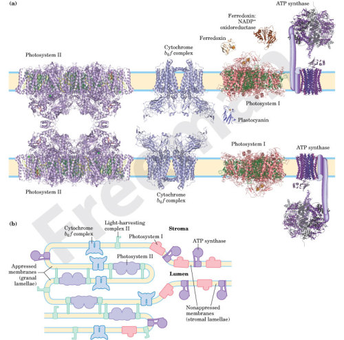
Membrane proteins of the thylakoid intercept light, they are then able to convert and store that as chemical energy.
mucholderthen: [ posted by currentsinbiology ] NCMIR...
[ posted by currentsinbiology ]
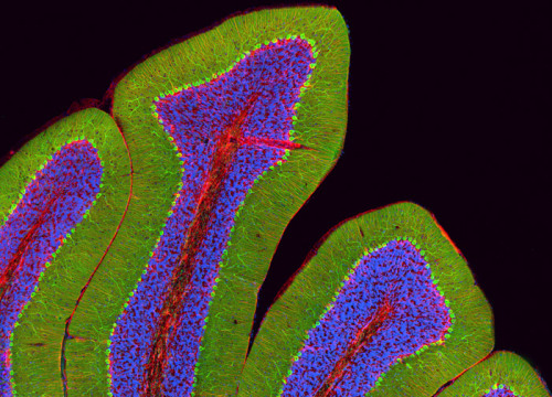
NCMIR microscopist Thomas Deerinck's image of a rat cerebellum. The image, a photomicrograph of a section of rat cerebellum, was achieved using Quantum Dot technology and multiphoton microscopy.
mucholderthen: CONVENTIONAL LIGHT MICROSCOPYSIM: Structured...
CONVENTIONAL LIGHT MICROSCOPY
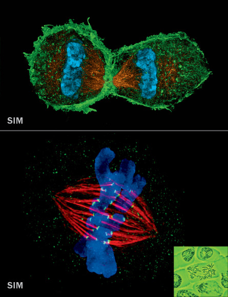
SIM: Structured illumination microscopy
Resolution: ~100 nm
- Structured illumination microscopy shines a striped pattern of light onto a sample.
- That light interacts with light from fluorescent tags on cellular material and generates a pattern of interference called a moiré fringe.
- Using a series of moiré fringes it's possible to mathematically extract and reconstruct a super-resolution image.
SIM is ideal for looking at entire cells in 3-D, ensembles of cells or multiple cellular structures at once.
_________________________PHOTO AT TOP: MITOSIS
- cellular division that produces two genetically identical daughter cells, is perhaps the most fundamental process in biology.
- After DNA replicates, the nuclear envelope surrounding it dissolves.
- Spindle fibers (gold above, red at right) align pairs of chromosomes (blue)
- then separate the genetic material into two daughter cells (shown forming).
Science News
June 15, 2013; Vol.183 #12Top: L. Schermelleh/Univ. of Oxford; Bottom: J. Stout/Indiana Univ.;
Bottom, right: Andrew Syred/Science Source(X)
The science notebook
The science notebook: explore new ideas .. discover science in a fun more intresting way :)mucholderthen: [ posted by txchnologist ] Earliest Stages of...
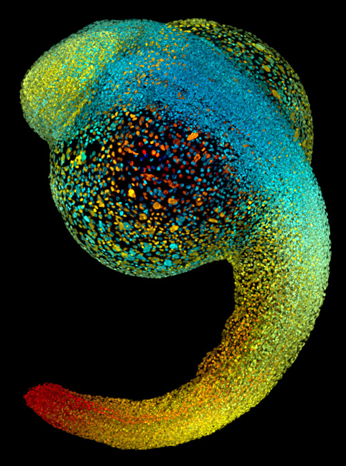
Image of the ~50,000 cell nuclei of a 22-hour-old zebrafish embryo. The fluorescently labeled cell nuclei are shown in a blue-to-red color code that indicates depth in the image.
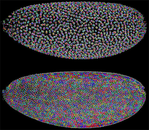
By following the color-coded cells of a Drosophila embryo (top) over time, each cell's lineage becomes trackable (bottom) with simultaneous multiview light sheet microscopy.
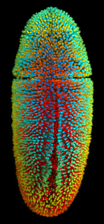
Image of the ~6,000 cell nuclei of a 3-hour-old fruit fly embryo. The fluorescently labeled cell nuclei are shown in a blue-to-red color code that indicates depth in the image.
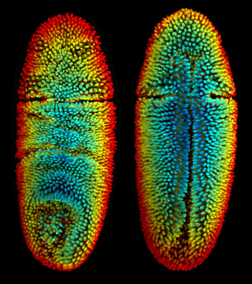
Dorsal and ventral views of a 3-hour-old fruit fly embryo. The embryo's ~6,000 fluorescently labeled cell nuclei are shown in a blue-to-red color code that indicates depth in the image.
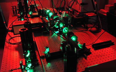
A SiMView microscope uses lasers to illuminate specimens while two cameras to the left and to the right of the central imaging chamber capture shots of the specimen's cells from different angles.
[ posted by txchnologist ]
Earliest Stages of Life Plumbed With Sheets of Laser Light
This month's issue of the journal Science focuses on advances in developmental biology, the study of how organisms grow and change from a single cell.
The edition includes a few stunning photographic examples from the lab of Philipp Keller at the Howard Hughes Medical Institute. Keller's team uses a new microscopy technique called simultaneous multiview light-sheet microscopy (SiMView), which created the detailed images of zebrafish and fruit fly embryos above. Click on the images to get a fuller description of what they depict.
currentsinbiology: Overcoming resistance to anti-cancer drugs...
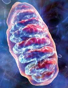
Overcoming resistance to anti-cancer drugs by targeting cell 'powerhouses'
Published: Wednesday, May 22, 2013 - 14:26 in Biology & NatureRe-routing anti-cancer drugs to the "power plants" that make energy to keep cells alive is a promising but long-neglected approach to preventing emergence of the drug-resistant forms of cancer — source of a serious medical problem, scientists are reporting. That's the conclusion of a new study published in the journal ACS Chemical Biology.
From e!Science News
sciencephotolibrary: Sperm production. Coloured scanning...

Sperm production. Coloured scanning electron micrograph (SEM) of sperm cells (spermatozoa) in the seminiferous tubules of the testis. This is the site of spermatogenesis (sperm production). Each sperm cell consists of a head (yellow), which contains the genetic material that fertilises the female egg cell, and a tail (blue), which propels the sperm. The heads of the sperm are buried in Sertoli cells (orange), which nourish the developing sperm.
Photo
emeraldmuse: moshita: IBB Blood Transfusion Packs is a...
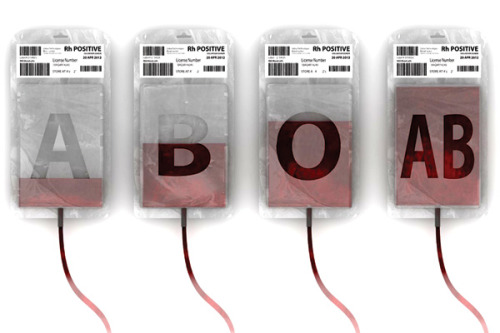

IBB Blood Transfusion Packs is a 2012 red dot award: design concept winner.
With the IBB Blood Transfusion Packs there will be no room for error while administering blood to those who need it. The packaging makes it almost impossible for you to make a mistake, because the letters A, B, or O appear prominently when the bag is filled with blood. Every part of the bags except the letters is translucent and this is what makes it distinctive.
The only thing better would be to have different male to female connectors on the IV lines going to the patents. That way it would be impossible to accidentally hook up the wrong blood type. I.e it would be like not being able to connect the wrong cable to a computer port.
Yes! great point!!!
moshita: IBB Blood Transfusion Packs is a 2012 red dot award:...
IBB Blood Transfusion Packs is a 2012 red dot award: design concept winner.
With the IBB Blood Transfusion Packs there will be no room for error while administering blood to those who need it. The packaging makes it almost impossible for you to make a mistake, because the letters A, B, or O appear prominently when the bag is filled with blood. Every part of the bags except the letters is translucent and this is what makes it distinctive.
frontal-cortex: Confocal image of a squid embryo. All nuclei...
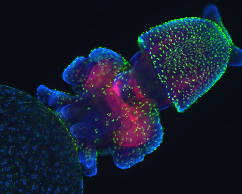
Confocal image of a squid embryo. All nuclei are stained with DAPI (blue). Phalloidin staining reveals neural structures (red), while cilia on the surface of the embryo are highlighted by acetylated tubulin staining (green). (Thenode.biologists.com)
lizardking90: Bit of Algae Credit: Frank Fox Fachhochschule...
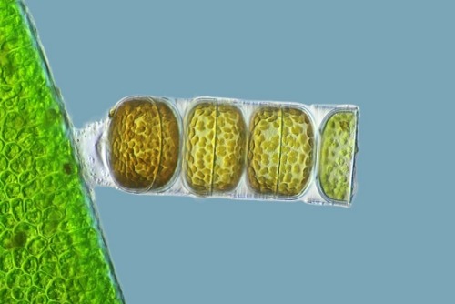
Bit of Algae
Credit: Frank Fox Fachhochschule Trier Trier, Rheinland-Pfalz, Germany
The third place winner of the 2011 Nikon Small World contest was this photo of living Melosira monliformis, a type of algae.
frontal-cortex: Fig. 4. Segregation patterns of two different...
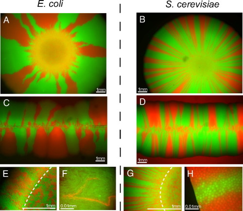
Fig. 4. Segregation patterns of two different microbial species are qualitatively similar but different in detail. (A and B) Both bacteria (E. coli) and yeast (S. cerevisiae) colonies exhibit spatial gene segregation when they grow colonies on agar plates. However, the number of (surviving) sectors is much larger in yeast colonies. For the linear inoculations (C and D), the Petri dish was gently touched with a sterile razor blade that was previously wetted by a liquid culture of a binary mixture of mutants. (E and G) Continuous patches of boundary regions and homeland (bounded by dashed line) at a magnification of ×51 for E. coli (E) and yeast (G). (F and H) Images at single-cell resolution (×100). (F) Tip of a sector dies out (E. coli). (H) Section boundary at the frontier (yeast).
frontal-cortex: Colonic dendritic cells (red) in crypts (SC)
Colonic dendritic cells (red) in crypts (SC)
abstractnerd: Science by BWJones on Flickr. An experiment
An experiment
ucsdhealthsciences: A scanning electron micrograph of ovarian...
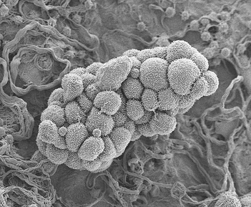
A scanning electron micrograph of ovarian cancer cells forming a small tumor. Image courtesy of the University of Gothenburg.
Potential New Way to Suppress Tumor Growth Discovered
Researchers at the University of California, San Diego School of Medicine, with colleagues at the University of Rochester Medical Center, have identified a new mechanism that appears to suppress tumor growth, opening the possibility of developing a new class of anti-cancer drugs.
Writing in this week's online Early Edition of the Proceedings of the National Academy of Sciences (PNAS), Willis X. Li, PhD, a professor in the Department of Medicine at UC San Diego, reports that a particular form of a signaling protein called STAT5A stabilizes the formation of heterochromatin (a form of chromosomal DNA), which in turn suppresses the ability of cancer cells to issue instructions to multiply and grow.
Specifically, Li and colleagues found that the unphosphorylated form of STAT promotes and stabilizes heterochromatin, which keeps DNA tightly packaged and inaccessible to transcription factors. "Therefore, genes 'buried' in heterochromatin are not expressed," explained Li.
Phosphorylation is a fundamental cellular function in which a phosphate group is added to a protein or molecule, causing it to turn it on or off or to alter its function. An unphosphorylated STAT lacks this phosphate group.
scienceyoucanlove: Meet your own symbionts: trillions of...
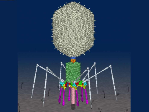
Meet your own symbionts: trillions of viruses
With deadly new viruses emerging these days in Saudi Arabia and China, it can be hard to imagine that viruses can be good for anything. It's easy to forget that we are home to trillions–perhaps quadrillions–of viruses on our healthiest days. And, according to a team of California scientists, those viruses are our symbiotic partners, creating a second immune system. These viruses serve as a defensive front-line, keeping bacteria from invading our gut lining and causing deadly infections.The viruses in question are far less familiar than, say, influenza viruses or Ebola viruses. They are known as bacteriophages, which means "eater of bacteria." And yet bacteriophages (or phages for short) are vastly more common than viruses that infect humans. They're more common than all the viruses that infect every animal on Earth. The reason is simple arithmetic: there are far more hosts for phages to multiply in than there are for viruses that infect our own cells. They're in the ground, in the oceans, under ice, and in the air. By some estimates, there are 1031 phages on Earth. That makes phages the most abundant life form, period.
Read more: http://phenomena.nationalgeographic.com/2013/05/20/meet-your-new-symbionts-several-trillion-viruses/
sagansense: (h) TROM - Chapter 1.2 Evolution of...
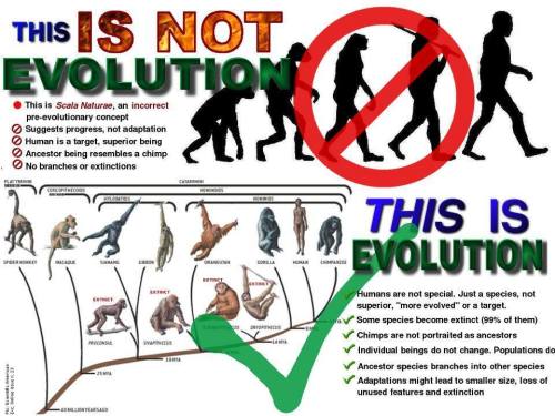
(h) TROM - Chapter 1.2 Evolution of Everything
TROM (The Reality of Me) represents the biggest documentary ever created, it is also the only one that tries to analyse everything : from science to the monetary system as well as real solutions to improve everyone's life. A new and ´real´ way to see the world.
The TROM documentary is trying to present, in a simplistic way, the world in which we, human beings, live. The world discovered so far, not some idea or personal choice. Moreover we tried to present alternative solutions to current problems and took into account the future, which promises to be more than interesting. An informative documentary, perhaps shocking and disturbing to many, depending on how you digest the information.
The documentary is divided into chapters and sub-chapters due to the documentary's excessive length (13 hours). Also all the parts are connected.
The documentary and website (tromsite.com/) are concentrated, so it is essential to read additional information which can be found in descriptions beside each section and the video player.
——————————-
I've published a post or two about this documentary before and although most of the information is already discussed or presented in various ways via multiple mediums on tumblr, youtube, webisodes, blogs, etc., I assure you, you haven't seen it delivered as professionally, and as clear & concise as this. The documentary is huge. I have the entire thing downloaded on an external hard drive as it's readily available for individual download at tromsite.com.
If there's one major documentary that should be viewed by everyone on this planet, this is it. Actually, the more I think about it, this documentary was 'one' of the major motivators for me to develop this blog as a means of publicly available access toward the promotion of scientific literacy. This documentary is what you've been looking for.
Enjoy, and share with someone, anyone, everyone. Introduce someone to reality as unveiled by the tools of science.
Over simplified, but a great place for learners. Enjoy the computer generated representation from the beginning of time to now…
currentsinbiology: A cluster of special nerve cells called...
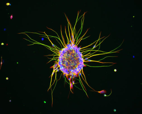
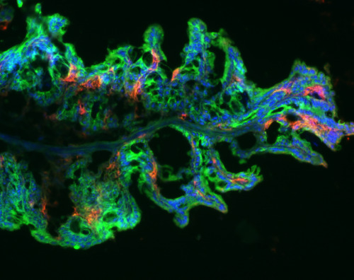
A cluster of special nerve cells called cerebellar granule cells, growing in culture. These cells naturally gather together, and when placed in a culture dish covered in a particular protein, they start sending out long projections (yellow/green) as they would in the developing brain.
Confocal micrograph by Ludovic Collin.
The above image shows a glass sculpture of a HIV particle. It...
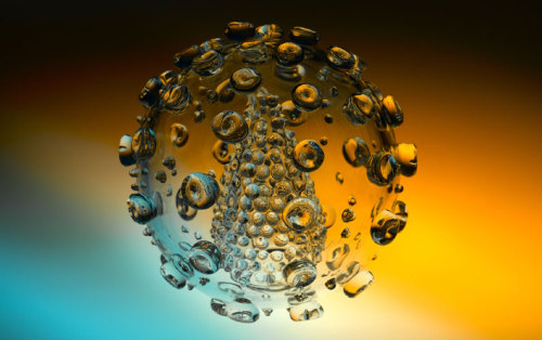
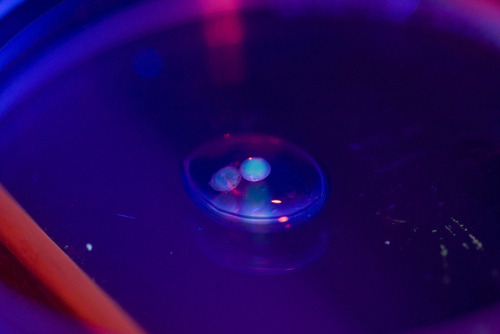
The above image shows a glass sculpture of a HIV particle. It was made by UK artist Luke Jerram, who received this letter in response from an anonymous fan in 2009:
Dear Luke,
I just saw a photo of your glass sculpture of HIV.
I can't stop looking at it. Knowing that millions of those guys are in me, and will be a part of me for the rest of my life. Your sculpture, even as a photo, has made HIV much more real for me than any photo or illustration I've ever seen. It's a very odd feeling seeing my enemy, and the eventual likely cause of my death, and finding it so beautiful.
Thankyou.
No hay comentarios:
Publicar un comentario